bluename
How to Treat Hibiscus Crown Gall: Remove Infected Tissue and Save Your Plant
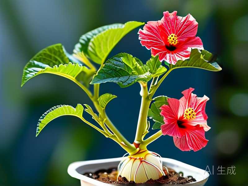
I’ll never forget the sinking feeling I had when I first noticed the rough, swollen lumps at the base of my prized hibiscus. What started as a small, corky bump had morphed into a grotesque, tumor-like growth. My initial panic led me down a rabbit hole of confusing advice. If you’re here, you likely know that dread. Crown gall, caused by the soil-borne bacterium Agrobacterium tumefaciens, can feel like a death sentence for your beloved plant. But through my own hands-on battle over several seasons, I’ve learned it’s not necessarily a terminal diagnosis. The most critical step is the surgical removal of the infected tissue. This guide is born from my personal experience—the successes, the mistakes, and the two-week recovery window that tells you if you’ve won the fight.
Understanding the Enemy: What is Hibiscus Crown Gall?
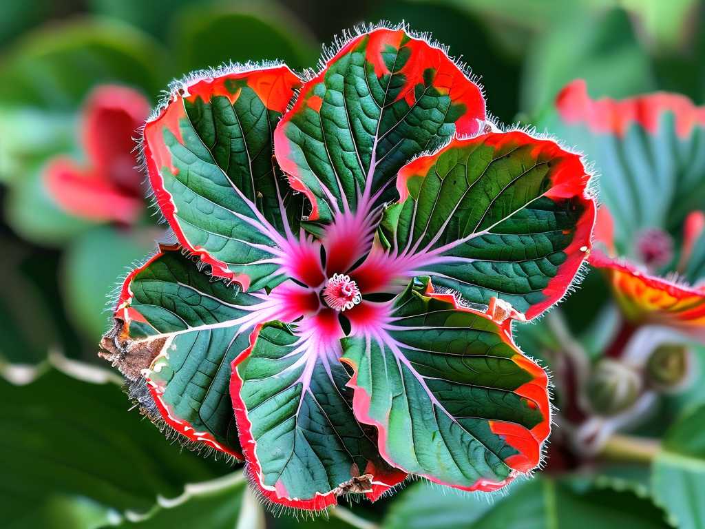
Before you can treat it, you must identify it correctly. Crown gall is not a fungal issue or physical damage; it’s a bacterial infection that tricks the plant into producing its own tumor. These galls typically form at the crown (soil line) but can also appear on stems and roots. They disrupt the vascular system, stunting growth, causing wilting, and eventually leading to plant death if left unchecked. According to the American Horticultural Society (AHS), this pathogen can persist in soil for years, making sanitation paramount. My first mistake was ignoring early signs, thinking it was just a weird scar. By the time I acted, the gall was the size of a golf ball.
My Step-by-Step Process for Surgical Removal
This isn’t a gentle pruning job; it’s a precise operation. I gathered my tools: a sharp, sterilized knife (I use a utility knife with replaceable blades), rubbing alcohol, a clean cloth, a sealant (I’ve used both grafting wax and a dedicated tree wound paint), a bucket of disinfectant solution (one part bleach to nine parts water), and heavy-duty gloves.
Step 1: Preparation and Sterilization I soaked my knife, pruning shears (for any follow-up trimming), and even my gloves in the bleach solution for a full five minutes. I then wiped them down with rubbing alcohol. Why such extremes? The AHS stresses that Agrobacterium spreads easily on contaminated tools. My early attempt using just wiped-down tools likely spread the bacteria to another plant—a hard lesson learned.
Step 2: The Removal Surgery On a dry, sunny morning, I carefully excavated the soil from around the crown to expose the full extent of the gall. The goal is to cut out all infected tissue, plus a margin of healthy-looking wood—about 1 to 2 inches beyond the gall. I made clean, smooth cuts, angling them slightly to shed water. I placed all removed tissue directly into a plastic bag, not letting it touch the soil. For a large, deep gall, this can look drastic, like you’re hollowing out the base. It’s frightening, but necessary.
Step 3: Disinfection and Sealing the Wound Immediately after cutting, I disinfected the wound. Some experts debate this step, but based on research from plant pathology resources, I applied a dilute bleach solution (10%) carefully to the cut surface with a small paintbrush, avoiding runoff onto roots. I let it air dry for 10 minutes. Then, I applied a thin layer of tree wound sealant. This protects the exposed tissue from secondary invaders while it calluses. I avoided thick, goopy applications that can trap moisture.
Step 4: Post-Op Care and Soil Management I removed and disposed of all the old mulch and soil from the area. I did not reuse it anywhere in the garden. I then watered the plant at the roots, avoiding the wound, and provided light shade for a few days to reduce stress. Crucially, I did not fertilize. The Royal Horticultural Society (RHS) advises against high-nitrogen fertilizers after crown gall surgery, as they can promote soft, susceptible growth.
The Critical 2-Week Observation Period
This is where patience is key. I marked my calendar and committed to daily, non-intrusive checks.
- Days 1-3: The plant looked shocked, with some slight wilting. This is normal. I ensured the soil stayed consistently moist but not soggy.
- Days 4-7: The wilting stabilized. My focus was on the wound edges. I looked for the first signs of callusing—a dry, smooth ridge forming around the cut. Any oozing, darkening, or softness would have been a bad sign.
- Days 8-14: By day 10, a definite callus ring had formed. No new, rough swellings appeared. A couple of small new leaves emerged from upper stems—the most hopeful sign! The two-week mark confirmed the initial surgery was successful; the plant was mobilizing its energy to heal and grow rather than feed new galls.
The Pitfalls I Encountered and How I Fixed Them
My journey wasn’t flawless. The first time, I didn’t cut widely enough. A tiny piece of infected tissue remained, and a new gall formed right next to the old scar within a month. I had to repeat the surgery, cutting even deeper. Another time, I over-watered in my anxiety, leading to root rot issues that compounded the stress. I learned to trust the process and check soil moisture with my finger. The most valuable fix was implementing a strict tool sterilization protocol between every single cut, not just at the start.
Preventing Future Crown Gall Infections
Treatment is only half the battle. Now, I always inspect new plants meticulously before purchase. I avoid planting hibiscus or other susceptible hosts (like roses and fruit trees) in a spot where gall has been found for at least two years. I boost overall plant health with balanced, slow-release fertilizer and proper watering to increase natural resistance. Creating well-draining soil conditions is a non-negotiable practice in my garden now.
Addressing Your Top Concerns
Can a hibiscus with crown gall ever fully recover? Yes, it can. “Recovery” means the plant survives, heals, and resumes healthy growth, but the bacterium may still be present in its system. The key is managing the disease to a point where it causes no harm. My oldest treated hibiscus has been gall-free for three growing seasons and blooms profusely, though I remain vigilant.
Is it safe to use the same tools on other plants after this? Absolutely not without thorough sterilization. After treating the gall, I disinfect my tools with bleach solution, then again with rubbing alcohol before they touch any other plant. I even keep a dedicated set of “surgery tools” for problematic cases.
Should I just remove and destroy the plant instead? For a severely compromised plant where galls encircle the entire crown or main roots, removal is the safest option to protect the rest of your garden. However, for localized galls on an otherwise valuable or healthy plant, surgical removal is a viable and often successful treatment. My decision tree is simple: if more than 1/3 of the crown circumference is affected, I consider removal.
Seeing that first new green shoot after the tense observation period is incredibly rewarding. Treating hibiscus crown gall through careful tissue removal is a direct, hands-on intervention that can save your plant. It requires courage, precision, and relentless sanitation. The process taught me more about plant physiology and resilience than any textbook could. By acting decisively at the first sign, sterilizing ruthlessly, and providing attentive aftercare, you can give your hibiscus a strong chance to thrive beyond this challenging disease. The fight is winnable.